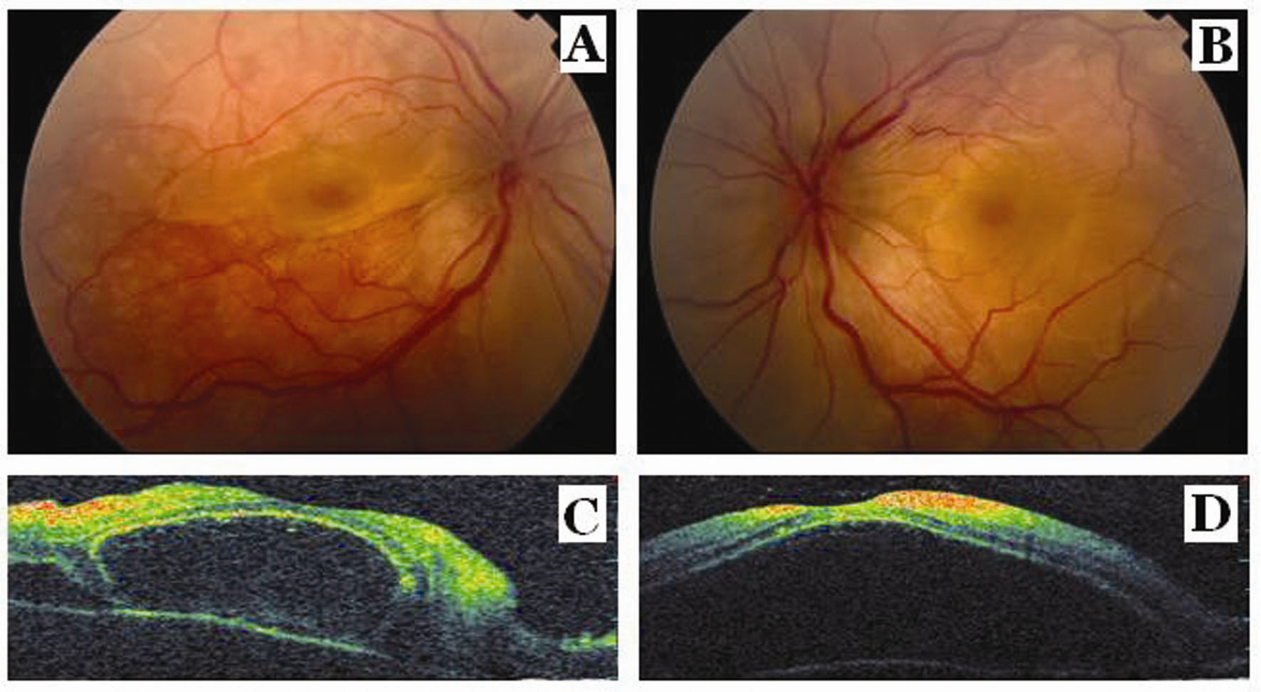
Figure 1.. Fundus photographs show serous macular detachment and retinal folds and optic disc edema in the right eye (A) and in the left eye (B). Optical coherence tomography scans show serous macular detachment with several subretinal septas in the right eye (C) and a giant serous macular detachment in the left eye (D) at the presentation.
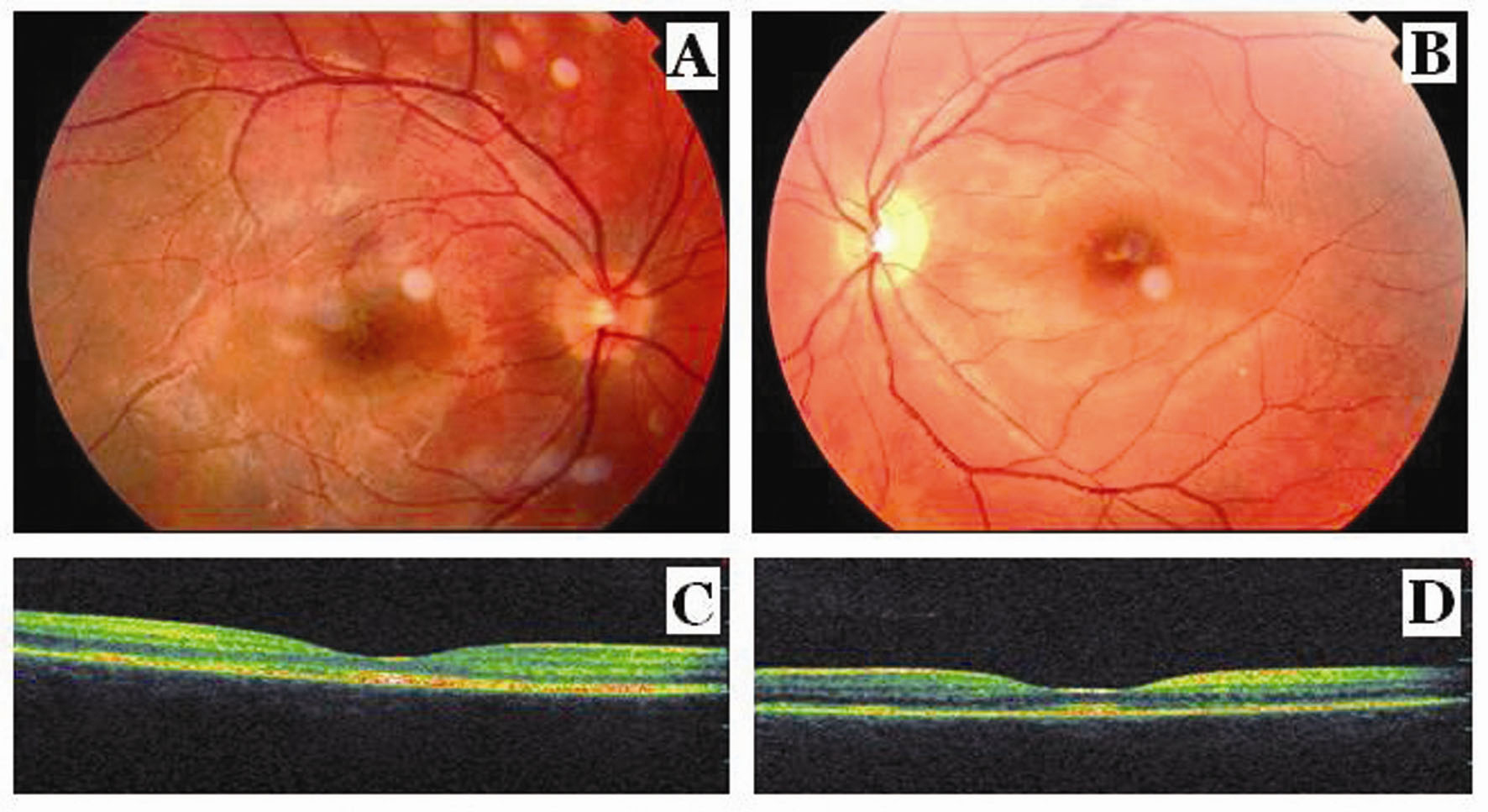
Figure 2.. Fundus photographs show complete reattachment of retina, retinal pigment epithelial changes as hyperpigmentation and fibrosis at the macula in the right eye (A) and complete reattachment of retina, retinal pigment epithelial changes as hyperpigmentation and fibrosis at the macula, and the pallor at the optic disk in the left eye (B) after the corticosteroid treatment. Optical coherence tomography scans show the resolution of macular detachment with formation of retinal pigment epithelial irregularities, epi macular membrane and sub foveal fibrosis in the right eye (C) and in the left eye (D) after the corticosteroid treatment.