Figures
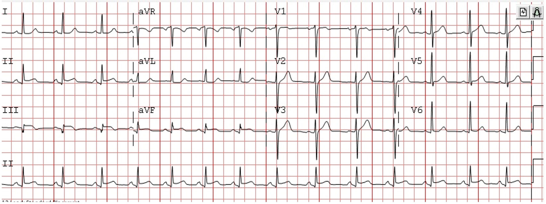
Figure 1. Electrocardiogram on admission demonstrating inferior ST elevation myocardial infarction.
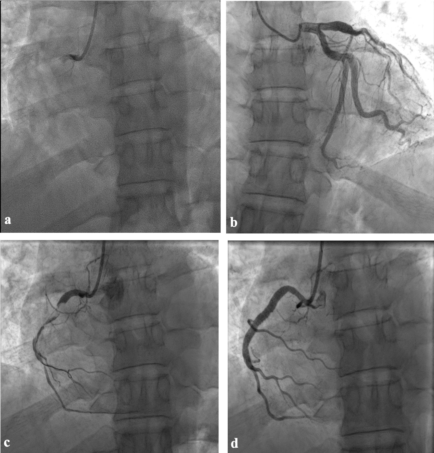
Figure 2. Coronary angiography and angioplasty. (a) Right coronary proximal total occlusion. (b) Left system ectasia in left anterior descending and first obtuse marginal arteries. (c) After thrombus aspiration attempt. (d) Final result after stent implantation.
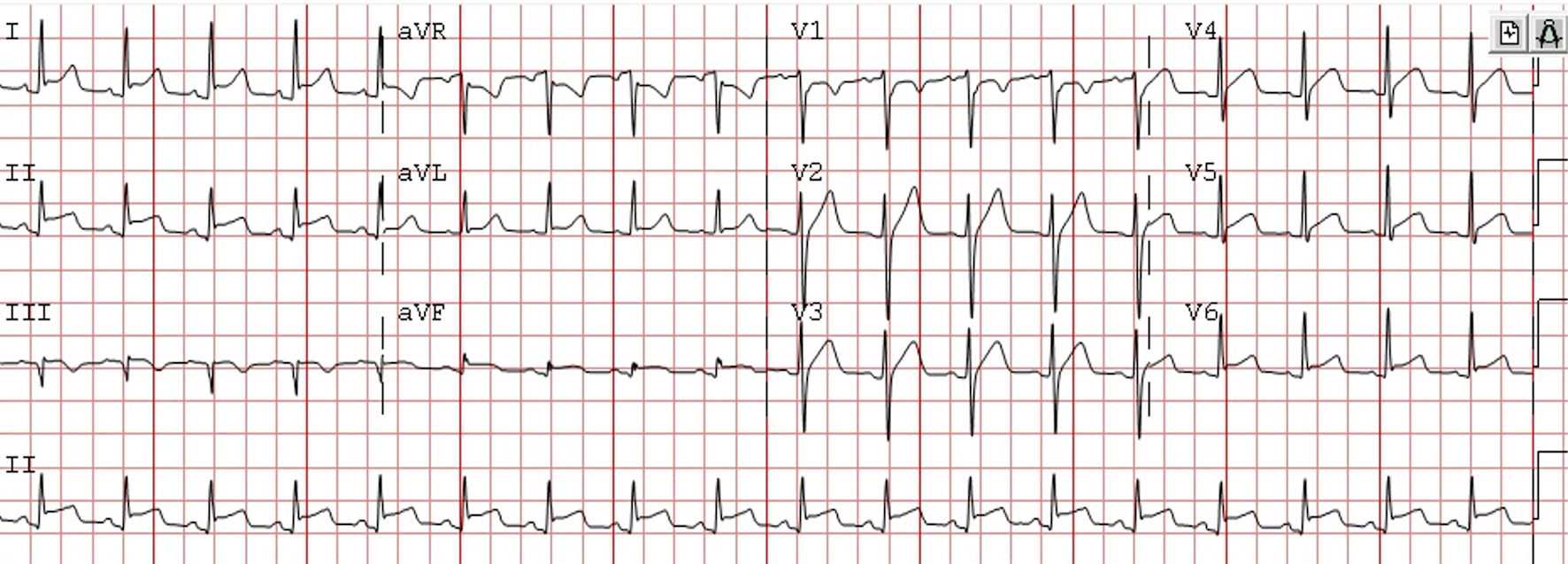
Figure 3. Electrocardiogram after 18 h in coronary care unit (CCU) demonstrating new widespread ST elevation more prominent in inferior leads II, III and aVF with ST depression in aVR.
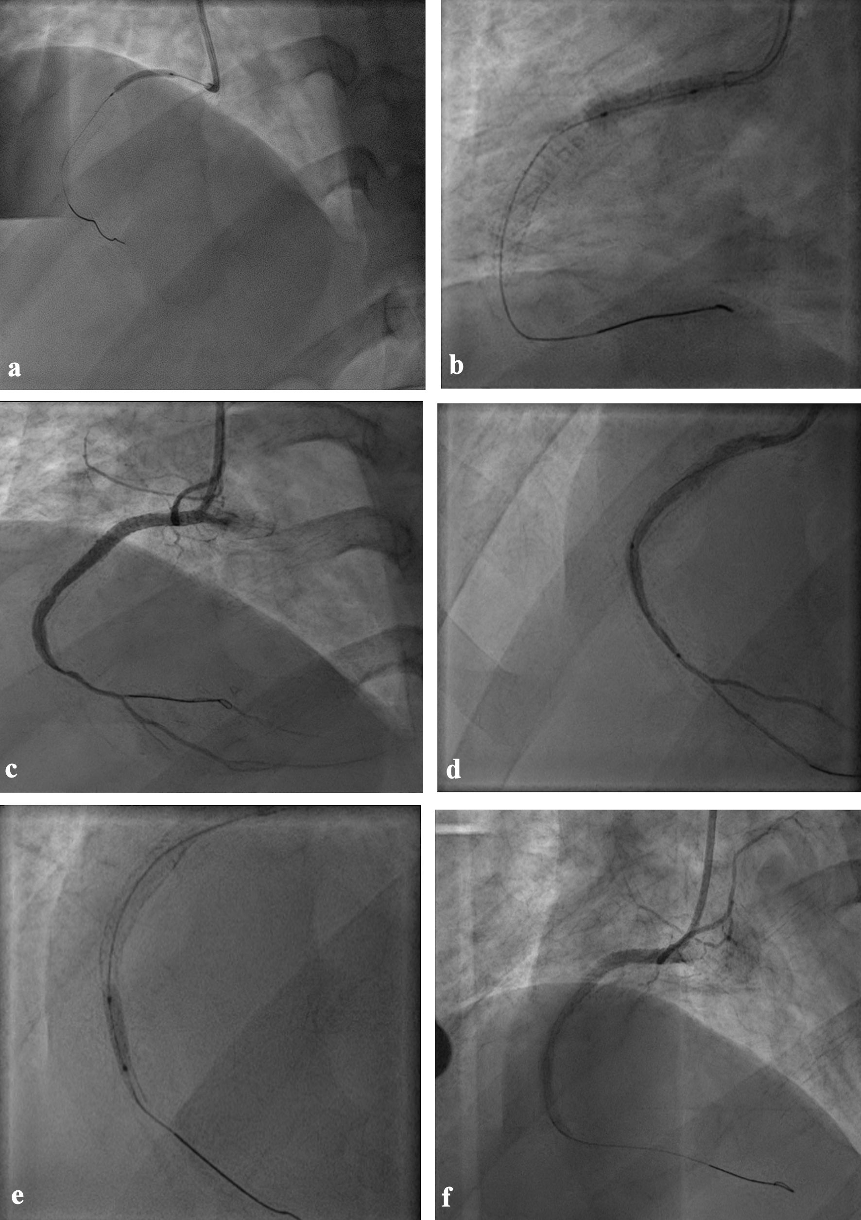
Figure 4. The second angioplasty (a) Balloon angioplasty after aspiration thrombectomy. (b) Stent implantation from ostium to mid-segment overlapping with the previous stent. (c) After injection of intracoronary adrenaline, adenosine and epinephrine. (d) Distal direct stent implantation. (e) Post dilatation using non-compliant balloon. (f) Final result after aspiration thrombectomy, vasodilator therapy and IVUS guidance to exclude mechanical complications showing distal no-reflow. IVUS: intravascular ultrasound.
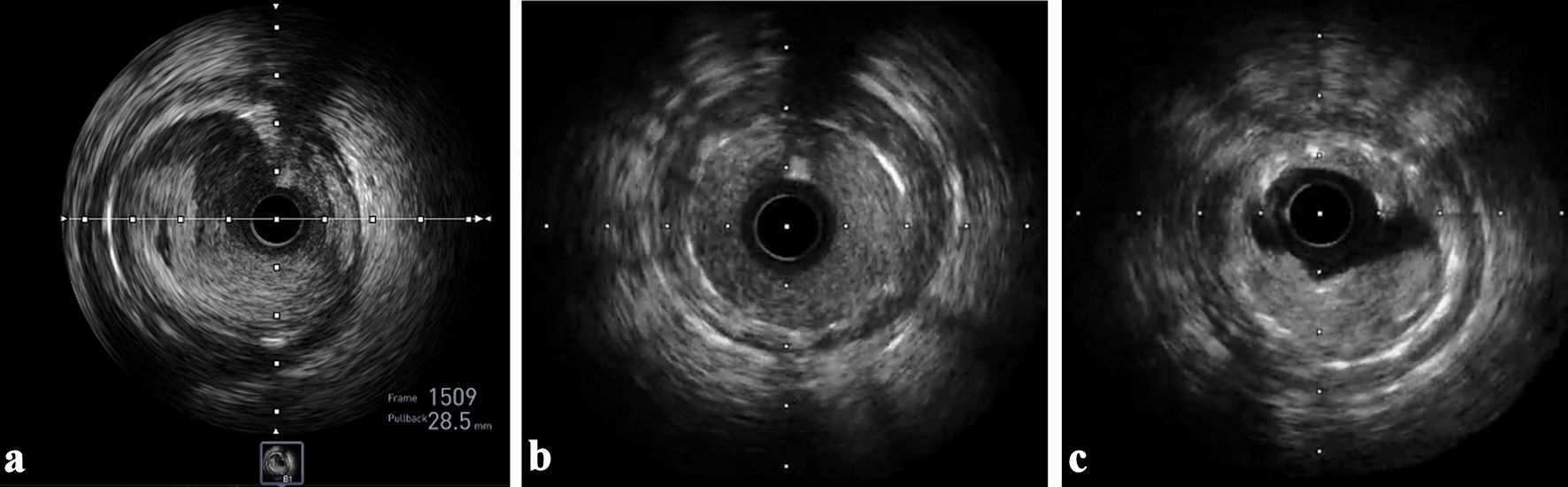
Figure 5. IVUS images. (a) Well opposed struts in proximal stent edge with no dissection. (b) Well opposed struts in distal stent edge with no dissection. (c) Acute in-stent thrombosis. IVUS: intravascular ultrasound.
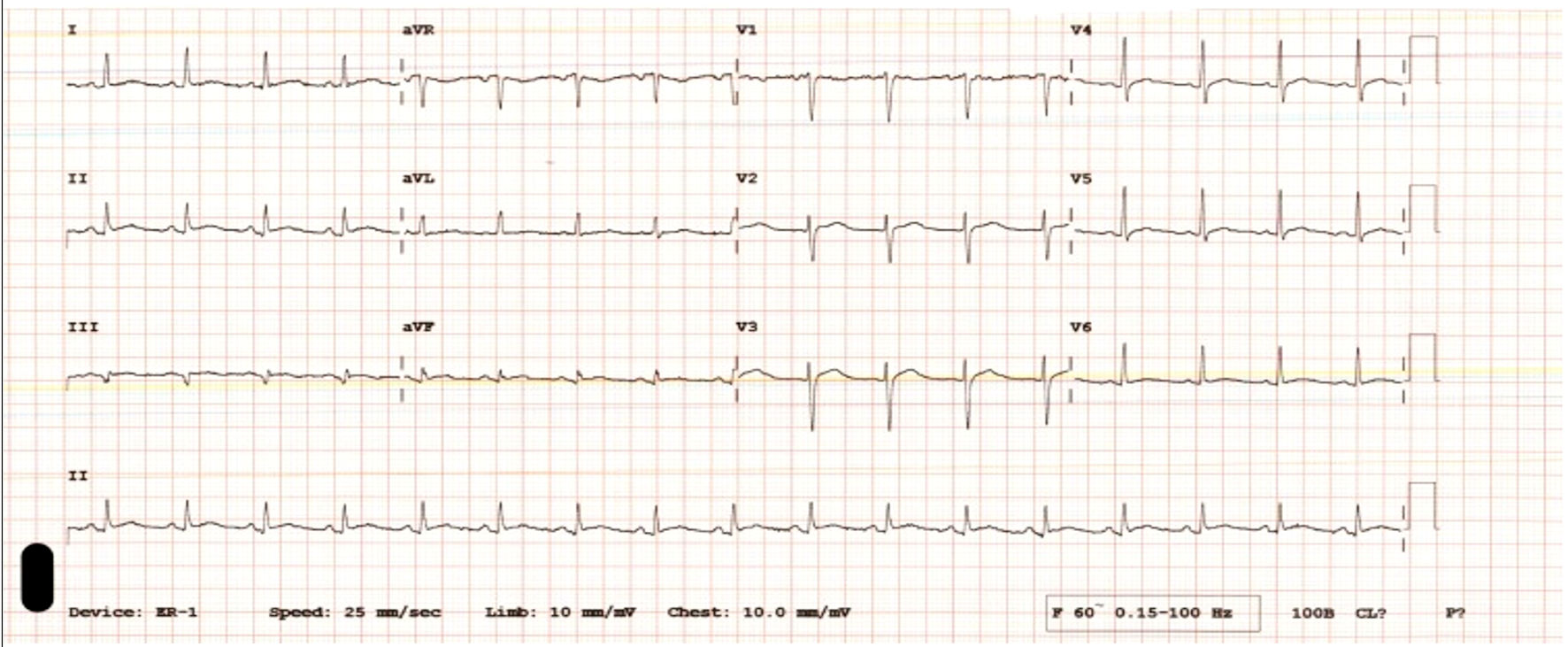
Figure 6. Electrocardiogram 4 days after discharge showing resolved wide-spread ST elevation.
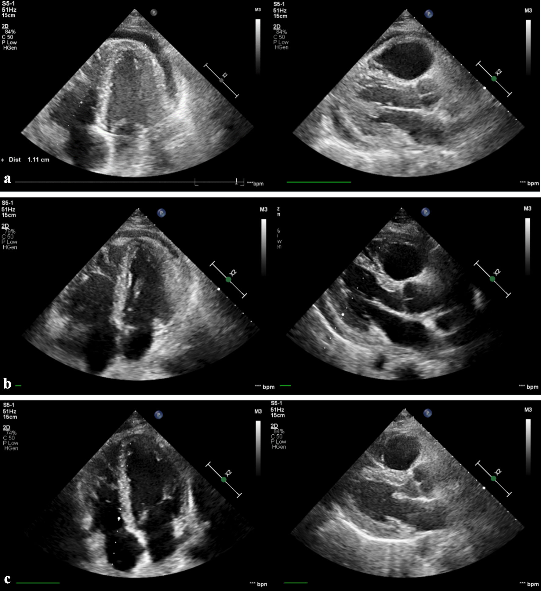
Figure 7. Echocardiography images. (a) Two weeks’ follow-up demonstrating moderate pericardial effusion at the apex. (b) Three weeks’ follow-up showing improvement to mild pericardial effusion. (c) Five weeks’ follow-up with complete resolution of pericardial effusion.
Table
Table 1. Inpatient Laboratory Investigations
| Parameter | Level | Normal range |
|---|
| HbA1c: hemoglobin A1c; LDL: low-density lipoprotein; HDL: high-density lipoprotein. |
| C-reactive protein (CRP) | 110 mg/L | 0 - 7 mg/L |
| HbA1c | 5.9% | 4-6% |
| Total cholesterol | 187 mg/dL | 140 - 200 mg/dL |
| LDL cholesterol | 116 mg/dL | 65 - 130 mg/dL |
| HDL cholesterol | 43 mg/dL | 35 - 86 mg/dL |
| Triglycerides | 140 mg/dL | 40 - 200 mg/dL |