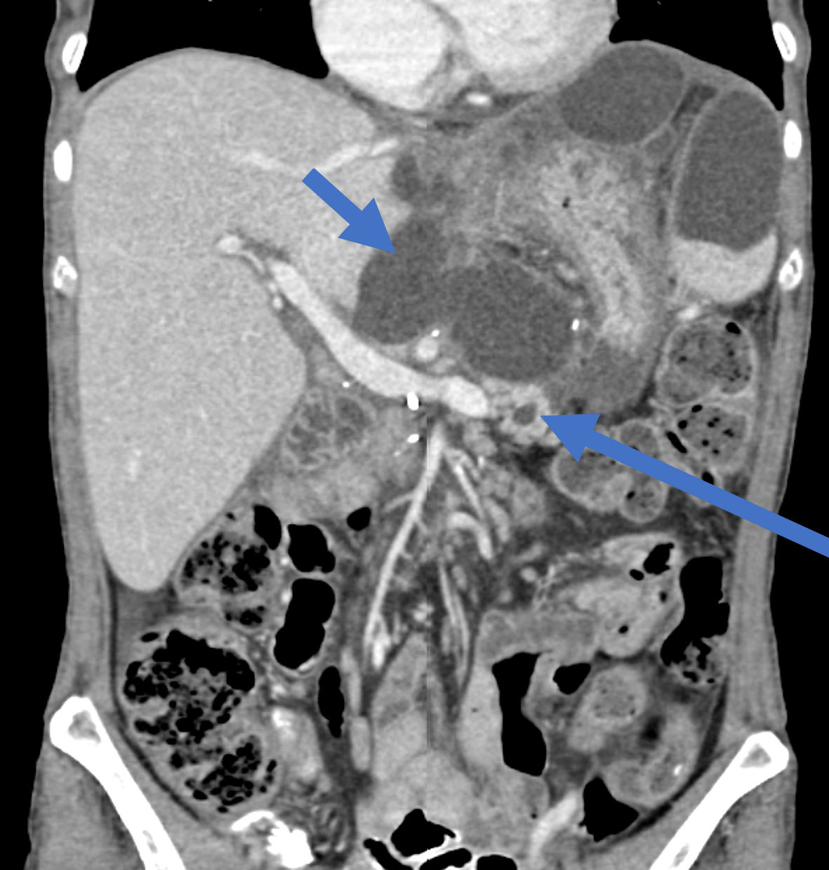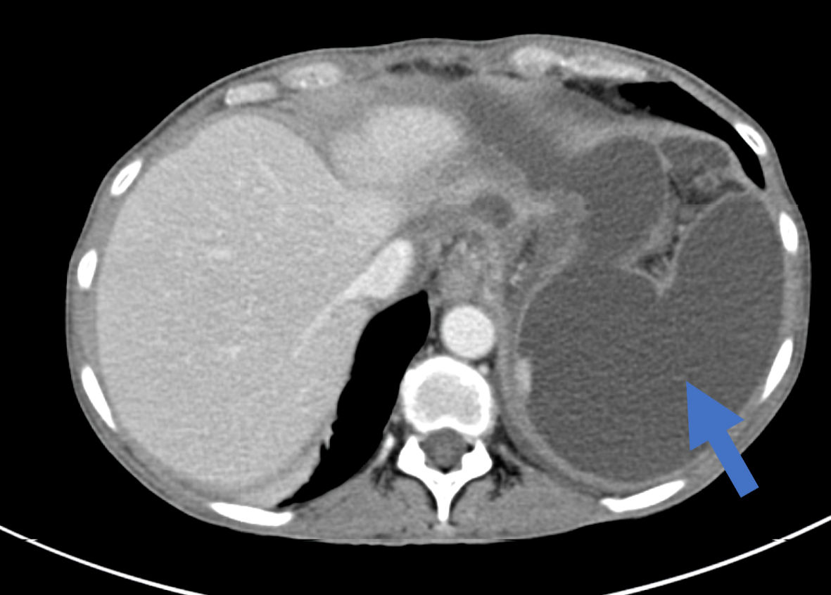
Figure 1. An axial abdominal CT scan showing a multilocular cystic intra-abdominal collection at the left upper abdomen (arrow). CT: computed tomography.
| Journal of Clinical Medicine Research, ISSN 1918-3003 print, 1918-3011 online, Open Access |
| Article copyright, the authors; Journal compilation copyright, J Clin Med Res and Elmer Press Inc |
| Journal website https://www.jocmr.org |
Case Report
Volume 16, Number 4, April 2024, pages 182-188
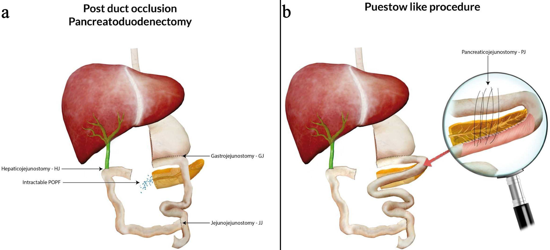
Intractable Postoperative Pancreatic Fistula: Undefined Type of Pancreatic Fistula Managed by Puestow-Like Procedure
Figures