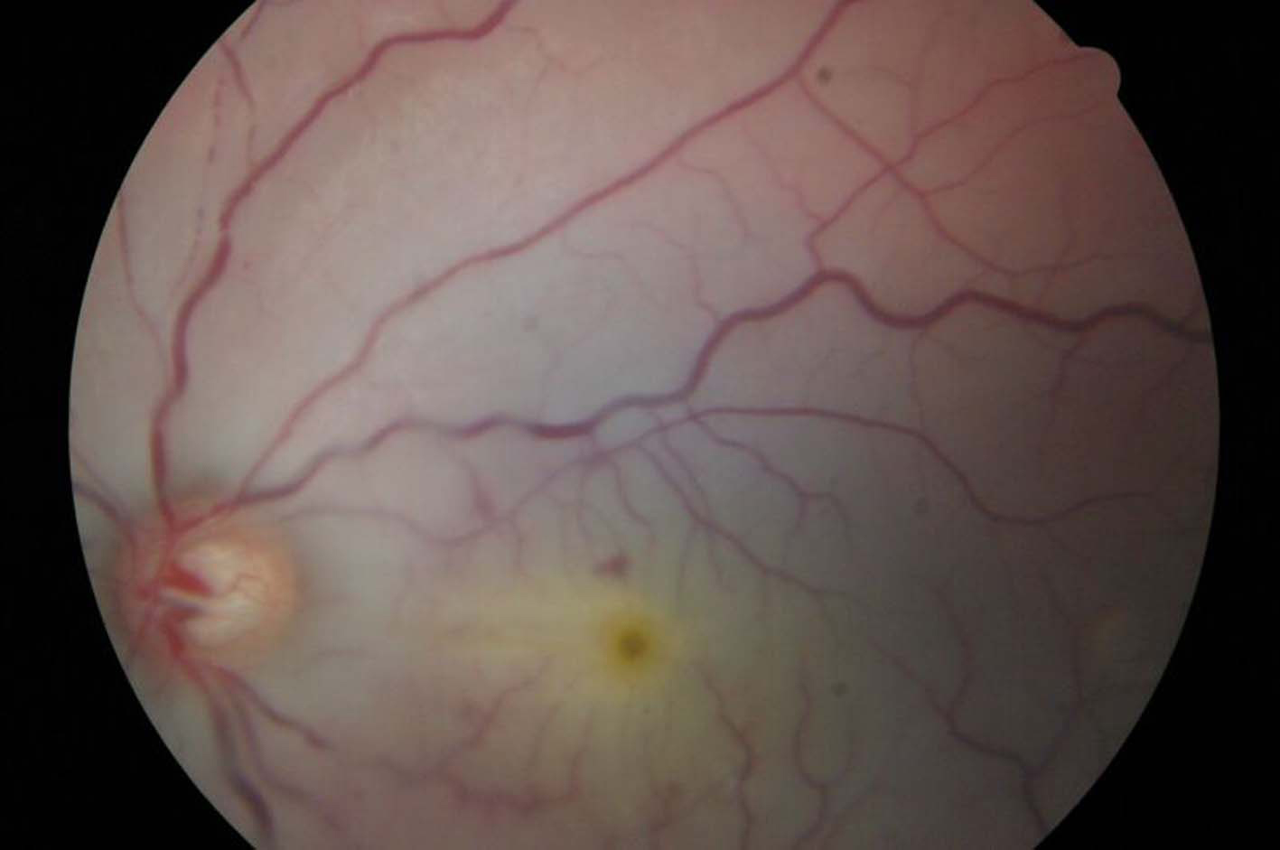
Figure 1.. Pale macular area with a cherry-red spot in the left eye.
| Journal of Clinical Medicine Research, ISSN 1918-3003 print, 1918-3011 online, Open Access |
| Article copyright, the authors; Journal compilation copyright, J Clin Med Res and Elmer Press Inc |
| Journal website http://www.jocmr.org |
Case Report
Volume 3, Number 1, February 2011, pages 55-57
Traumatic Optic Neuropathy and Central Retinal Artery Occlusion Following Blunt Ocular Trauma
Figures