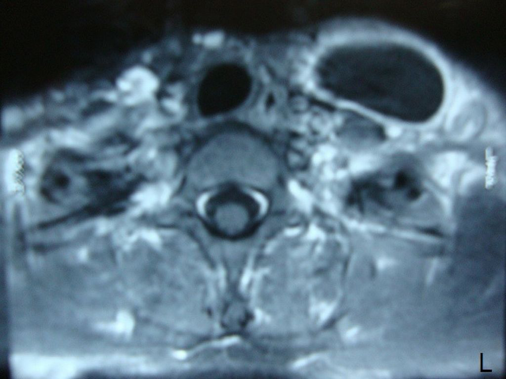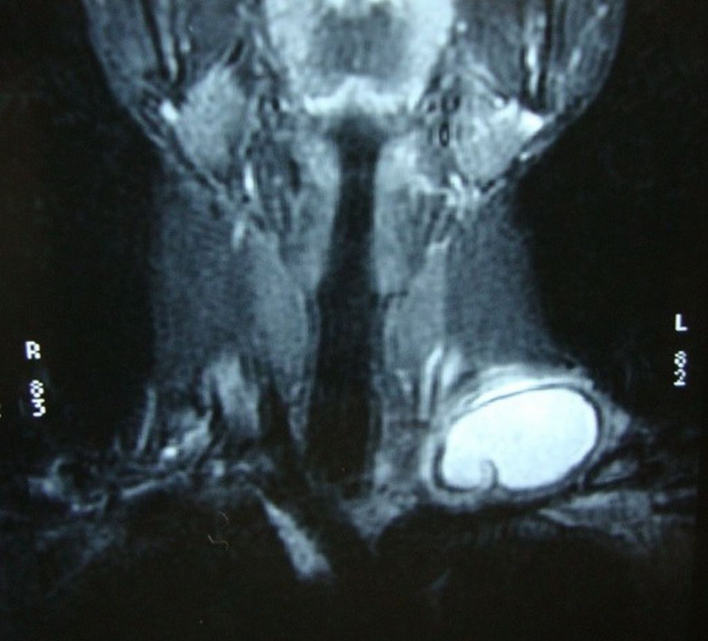
Figure 1.. Coronal T2-weighted MRI showing a unilocular, well-defined and high signal intensity cystic lesion with hypointense rim.
| Journal of Clinical Medicine Research, ISSN 1918-3003 print, 1918-3011 online, Open Access |
| Article copyright, the authors; Journal compilation copyright, J Clin Med Res and Elmer Press Inc |
| Journal website http://www.jocmr.org |
Case Report
Volume 3, Number 1, February 2011, pages 52-54
Primary Hydatid Cyst: An Unusual Cause of a Mass in the Supraclavicular Region of the Neck
Figures