Journal of Clinical Medicine Research, ISSN 1918-3003 print, 1918-3011 online, Open Access
Article copyright, the authors; Journal compilation copyright, J Clin Med Res and Elmer Press Inc
Journal website https://www.jocmr.org

Case Report

Volume 15, Number 7, July 2023, pages 384-389


Histopathological Findings of Ectopic Pregnancy in Contraceptive-Wearing Woman

Figures

Figure 1.
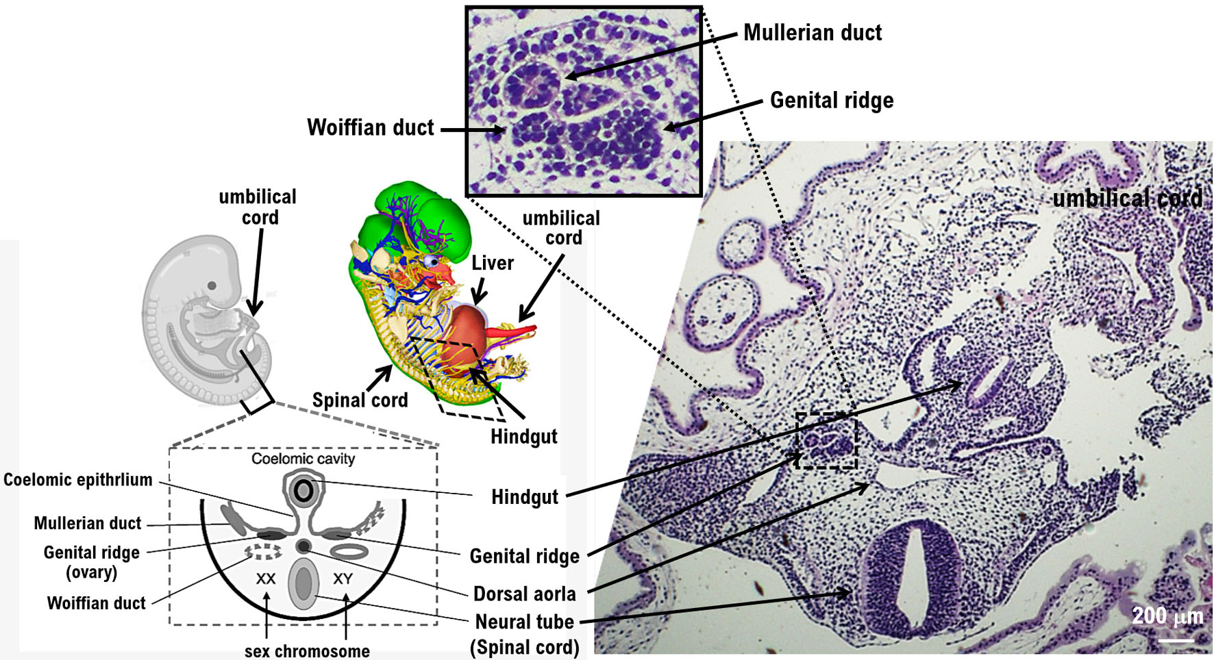
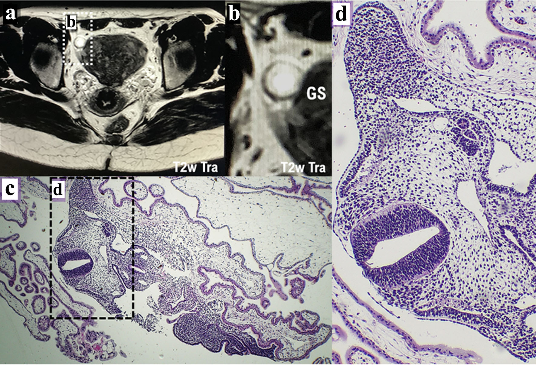
Figure 1. Diagnosis of ectopic pregnancy by contrast-enhanced MRI and histopathological examination. (a) The result described from contrast-enhanced MRI examination (T2w Tra) shows the possibility of ectopic pregnancy. The white dotted area shown in a is presented as panel b. (b) The GS in the right fallopian tube is indicated by a white spherical structure. Ectopic pregnancy was suspected because MRI showed a cystic lesion with ring-shaped diffusion restriction and contrast enhancement in the right fallopian tube. (c) A specimen of the resected right fallopian tube was used for pathological diagnosis. The black dotted area shown in panel c is presented as panel d. (d) Histopathological examination reveals the histology of the developing fetus during the growth period. GS: gestational sac; MRI: magnetic resonance imaging.
Figure 2.
Figure 2. Structure of various tissues of the growing fetus via histopathological examination. Histopathological examination reveals the structure of various tissues of the growing fetus after the ninth week of pregnancy. The images show the development of the neural tube (spinal cord), hindgut, and dorsal aorta. Pathological examination shows the development of the genital ridge. The genital ridge (or gonadal ridge) is the precursor to the gonads. Initially, the genital ridge consists mainly of mesenchyme and cells of underlying mesonephric origin. Once oogonia enter this area, they attempt to associate with these somatic cells. Pathological results suggest that the fetus may be female.