Journal of Clinical Medicine Research, ISSN 1918-3003 print, 1918-3011 online, Open Access
Article copyright, the authors; Journal compilation copyright, J Clin Med Res and Elmer Press Inc
Journal website https://www.jocmr.org

Review

Volume 13, Number 7, July 2021, pages 367-376


The Role of Cervical Muscles Morphology in the Surgical Treatment of Degenerative Disc Disease: Clinical Correlations Based on Magnetic Resonance Imaging Studies

Figure

Figure 1.
Figure 1. Axial T2 MRI image that demonstrates the muscle layers of the cervical spine of a 40-year-old asymptomatic woman at C5/C6 level. MRI: magnetic resonance imaging; LCA: longissimus capitis; LC: longus colli; LS: levator scapulae; MU: multifidus; SCA: scalenus anterior; SC: splenius capitis; SMC: semispinalis capitis; SCM: sternocleidomastoid; SMCe: semispinalis cervicis; T: trapezius.

Tables

Table 1. MRI-Based Studies of Cervical Spine Musculature of Patients That Underwent an Anterior Approach for Degenerative Disc Disease
 
Authors/year of publicationType of studyNumber of patientsType of surgeryConclusions
MRI: magnetic resonance imaging; ACDF: anterior cervical discectomy and fusion; VBA: vertebral body area; ASD: adjacent segment degeneration.
Choi et al, 2016 [19]Retrospective case-control study243 divided in fusion and no fusion groupsACDFIncreased dimensions of extensor semispinalis cervicis at C5/C6 level was found positively correlated with a successful and quicker fusion process.
Matsumoto et al, 2012 [20]Prospective case-control study31 patients that were operated with ACDF approach, and 32 healthy adults as control groupACDFPosterior cervical muscles were not different significantly between the two groups with only a small decrease in the cross-sectional area (CSA) in the ACDF group. These changes were not significantly correlated to pain or functionality scores.
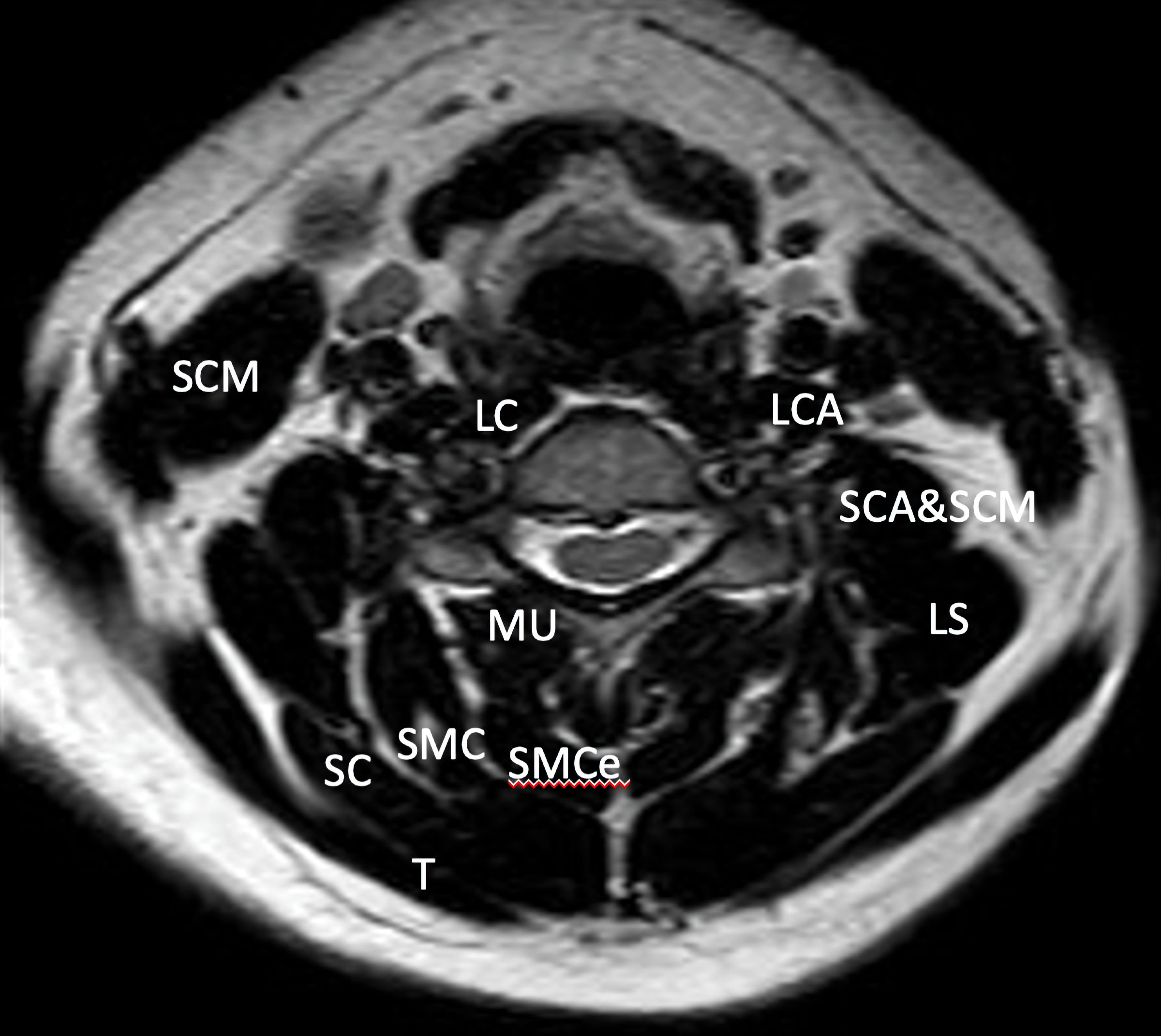
Thakar et al, 2014 [21]Retrospective case-control study67 patients with cervical spondylotic myelopathy and normal subjectsAnterior cervical corpectomy-fusion-platingNormal population has significant higher ratios of CSAs of the superficial, deep flexor (DF), and deep extensor (DE) paraspinal muscles than patients’ group (P < 0.001). Higher age patients and female patients had a lower total extensor CSA/VBA ratio (P < 0.001), while a longer duration of symptoms significantly predicted a greater total flexor/total extensor CSA ratio (P = 0.02). Among the muscle area ratios, the DF/DE ratio demonstrated a negative correlation with segmental angle change in the subgroup with preoperative straight or kyphotic segmental angles (P = 0.04 in the single corpectomy group, P = 0.01 in the two-level corpectomy group). There was no correlation of any of the muscle ratios with change in Nurick grade.
Thakar et al, 2019 [22]Prospective study45 patients with single level radiculopathy-myelopathy or myelopathyACDFDeep flexors areas as well as the ratio of deep flexors areas to deep extensors areas have a significant prognostic value of Nurick grade improvement.
Wong et al, 2020 [23]Retrospective case-control study32 patients with adjacent level disease after a two-level ACDF procedure; 30 patients that did not have this complicationACDFAsymmetry of fat at C5 (coefficient: 0.06), standardized measures of C7 lean (coefficient: 0.05) and total CSA measures (coefficient: 0.05) were the strongest predictors of early onset ASD.


Table 2. MRI-Based Studies of Cervical Spine Musculature of Patients That Underwent a Posterior Approach for Degenerative Disc Disease
 
Authors/year of publicationType of studyNumber of patientsType of surgeryConclusions
MRI: magnetic resonance imaging; CSA: cross-sectional area; QOL: quality of life; CSM: cervical spondylotic myelopathy.
Ashana et al, 2017 [24]Retrospective study6118 laminoplasty and 43 laminectomy and fusion patientsAuthors found a 2.19 times increase of the degree of atrophy in patients who sustained laminectomy and fusion. This study concluded that paraspinal muscle atrophy is correlated to anatomically larger surgical procedures.
Chen et al, 2019 [25]Retrospective study6336 patients with laminoplasty and posterior muscle-ligament complex preservation, and 27 patients with traditional laminoplastyThe preservation of posterior muscle-ligament complex protects patients from a postoperative muscle volume decrease which was noted in the traditional laminoplasty patients’ group (P < 0.01).
Kim et al, 2020 [26]Retrospective study106Cervical laminoplastyHigher T1 slope and less regional cross-sectional areas at C7 - T1 level were associated with loss of cervical lordosis.
Kotani et al, 2009 [27]Retrospective study84conventional open-door laminoplasty and deep extensor muscle-preserving laminoplastyLaminoplasty with deep extensor muscle-preserving approach appeared to be effective in reducing the axial pain and deep muscle atrophy, as well as improving cervical spine function and QOL when compared to conventional open-door laminoplasty.
Kotani et al, 2012 [28]Prospective study90Conventional open-door laminoplasty and deep extensor muscle-preserving laminoplastyThere is a superiority of deep extensor muscle-preserving laminoplasty in terms of postoperative axial pain, QOL, and prevention of atrophy of the deep extensor muscles over conventional open-door laminoplasty for the treatment of CSM.
Lee et al, 2018 [29]Prospective study144Laminoplasty procedureStatistical analysis revealed that high T1 slope and low summation of CSAs at each level of the semispinalis cervicis is correlated with loss of cervical lordosis. The level of the semispinalis cervicis associated with greater loss of lordosis was the C6 level.


Table 3. Classification of Cervical Spine Muscles Based on Their Function
 
Function of cervical musculature
  Flexion
    Sternocleidomastoid
    Longus colli
    Longus capitis
    Rectus capitis anterior
    Stylohyoid
    Digastric
    Mylohyoid
    Geniohyoid
    Omohyoid
    Sternohyoid
    Sternothyroid
  Extension
    Splenius capitis
    Splenius cervicis
    Semispinalis capitis
    Semispinalis cervicis
    Longissimus capitis
    Longissimus cervicis
    Trapezius
    Interspinal
    Rectus capitis posterior major
    Rectus capitis posterior minor
    Obliquus capitis superior
  Rotation
    Sternocleidomastoid
    Trapezius
    Splenius capitis
    Longissimus capitis
    Obliquus capitis inferior
    Rectus capitis posterior major
    Longus capitis
    Rotators
    Multifidus
  Lateral flexion
    Scalene
    Splenius capitis
    Longus colli
    Levator scapulae
    Iliocostalis cervicis
    Intertransversarii