Figures
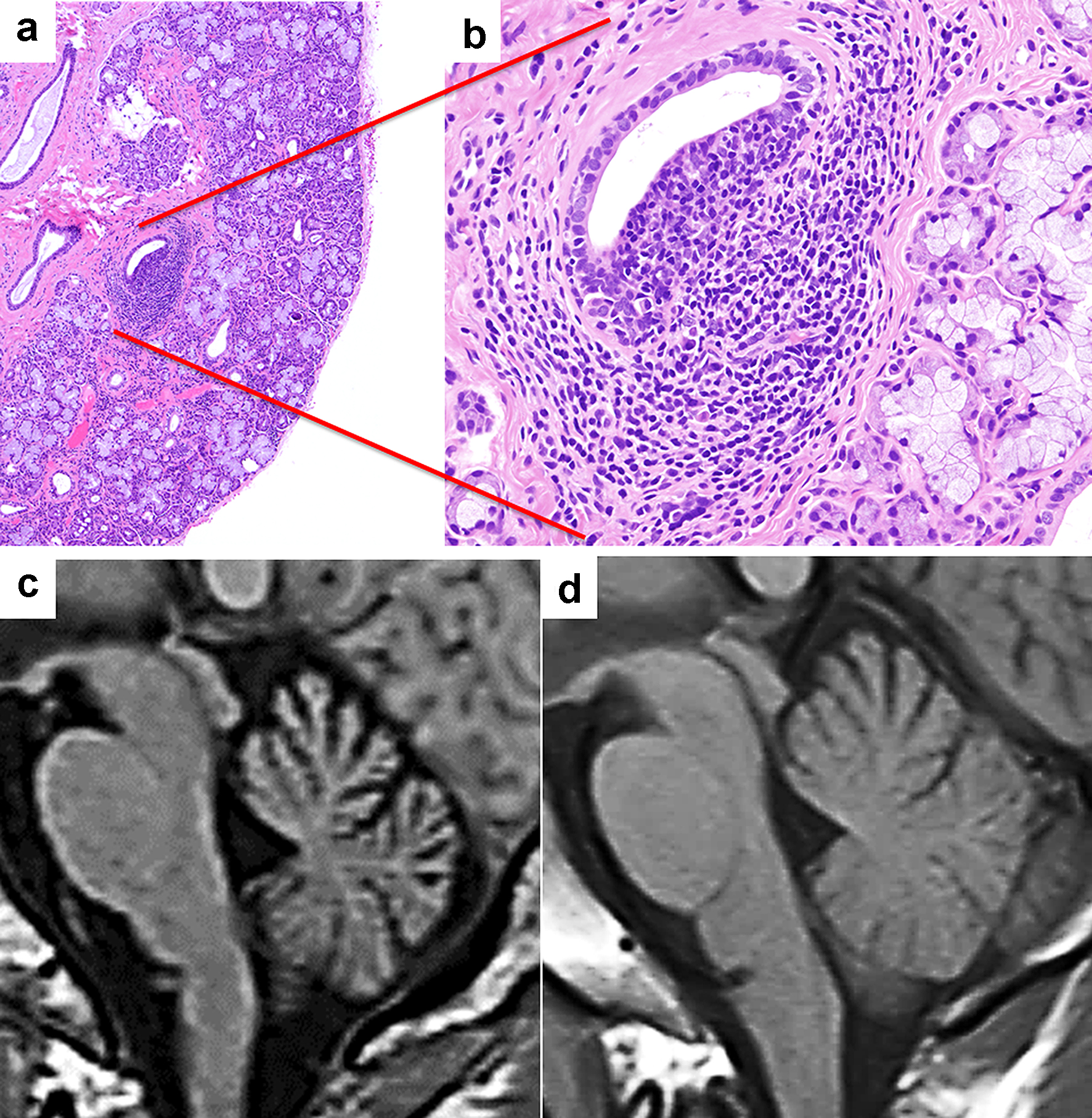
Figure 1. Patient 1 (Sjogren’s syndrome with cerebellar degeneration). (a, b) Photomicrographs of a salivary gland tissue section showing mild lymphocytic infiltration between the acini and glands (hematoxylin and eosin stain, magnification for a: × 40, b: × 200). (c, d) Sagittal views of the brain using magnetic resonance imaging T1 sequences. (c) The MRI performed 1 year after the initial presentation revealed cerebellar atrophy. (d) The initial MRI was unremarkable.
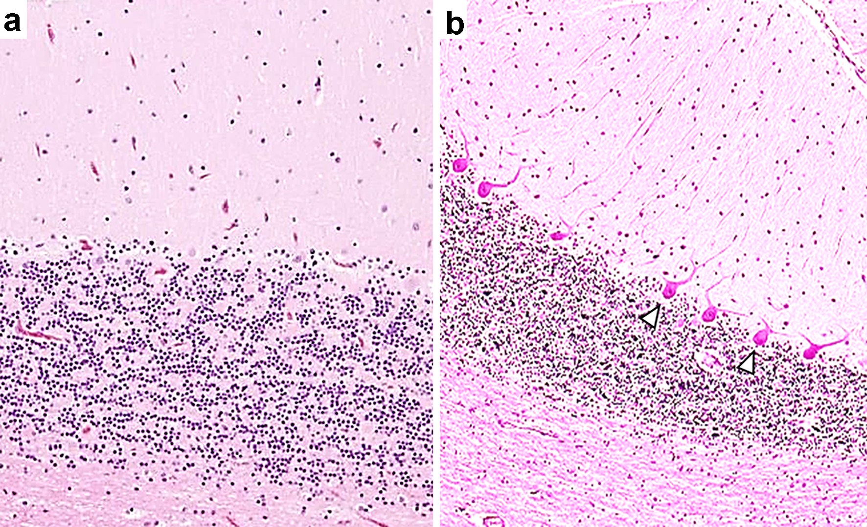
Figure 2. A selective loss of Purkinje cells in a patient with Sjogren’s syndrome with cerebellar degeneration. The patient was an 84-year-old woman who presented with nystagmus, dysarthria and ataxia in all limbs. Anti-Ro/SSA antibodies concentrations were determined to be > 500 U/mL, and anti-La/SSB antibodies were 41.1 U/mL. Intravenous immunoglobulin therapy was moderately effective. The patient died of pneumonia at age 85 years; an autopsy was then performed [6]. (a) A photomicrograph shows a mild-to-moderate decrease in the number of Purkinje cells in association with mild Bergman gliosis elsewhere in the cerebellar cortex. (b) This photomicrograph shows a normal human cerebellar tissue section with an appropriate number of Purkinje cells. Hematoxylin and eosin stain. The photomicrographs of tissue sections from this patient were reprinted and modified with permission from the publisher ([6], figure 4, Creative Commons CC BY). Arrowheads indicate Purkinje cells.
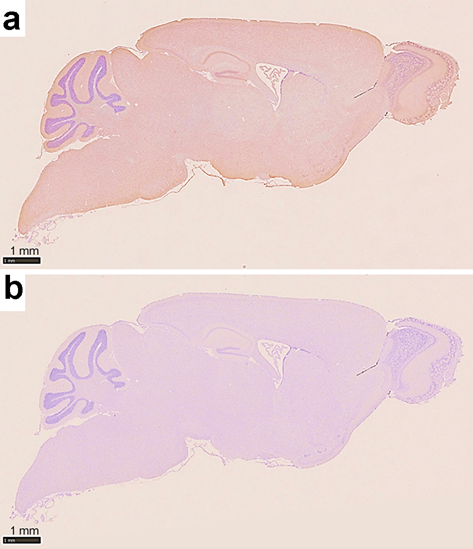
Figure 3. Photomicrographs of immunohistochemistry of a sagittal section of the murine brain. (a) The section is stained with a commercially available anti-Ro52/TRIM21 antibody showing Ro52/TRIM21 expression throughout the entire brain, including the hippocampus, cerebral cortex and cerebellum. (b) Negative control using an unrelated control antibody (anti-rabbit Ig) showing a lack of background staining (counterstain: hematoxylin).
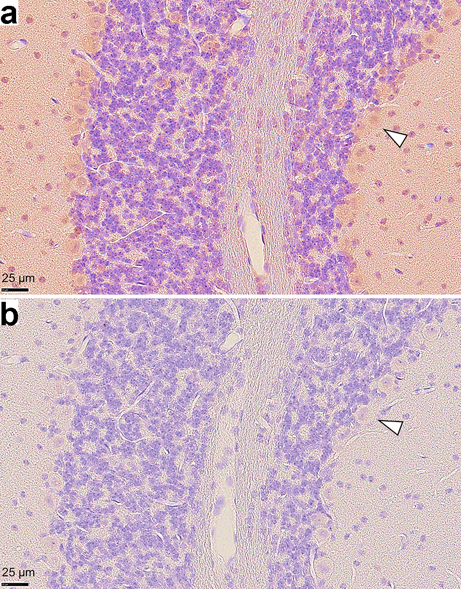
Figure 4. Photomicrographs of immunohistochemistry on murine cerebellar tissue sections. (a) Ro52/TRIM21 expression is shown using an anti-Ro52/TRIM21 antibody, but (b) is not present after staining with an anti-rabbit Ig (negative control). High Ro52/TRIM21 expression was observed in the cytoplasm of Purkinje cells. Arrowheads indicate representative Purkinje cells (magnification: × 40, counterstain: hematoxylin).
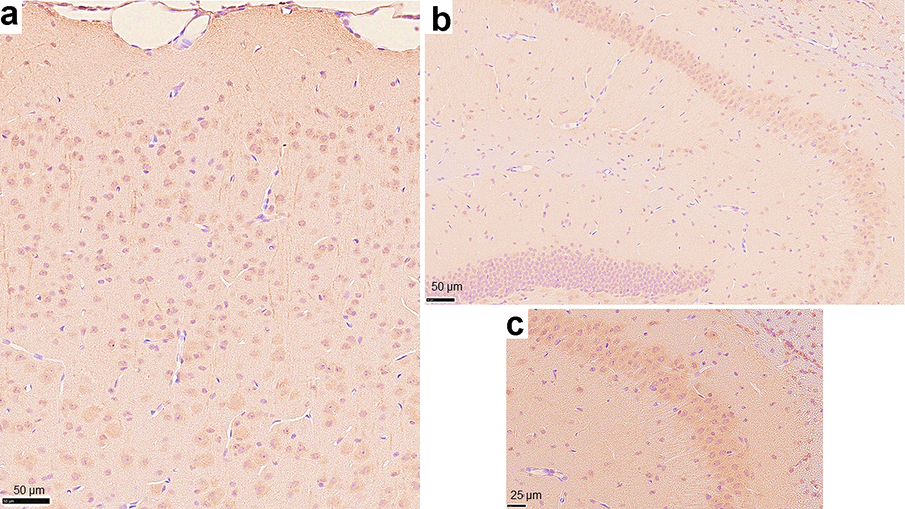
Figure 5. Photomicrographs of immunohistochemistry on other regions of the murine brain using the anti-Ro52/TRIM21 antibody. Cells that stained positively with the anti-Ro52/TRIM21 antibody were observed throughout (a) the cerebral cortex and (b) the hippocampus (magnification: × 20). (c) High magnification image representing the CA3 region of the hippocampus (magnification: × 40, counterstain: hematoxylin).
Table
Table 1. The Clinical Manifestations of Sjogren Syndrome Patients With Cerebellar Degeneration From Our Literature Review
| Author | Year/sex/age | Dry eye/mouth | Labial gland biopsy | ANA | Anti-Ro/SSA | Anti-La/SSB | Ataxia | Nystagmus | Dysarthria | Cerebellar atrophy | Previous diagnosis of SjSa | Therapy | Prognosis |
|---|
| aWhether the diagnosis of SjS had been made prior to the onset of cerebellar degeneration. SjS: Sjogren’s syndrome; M: male; F: female; NP: not performed; ANA: antinuclear antibody; Pre: prednisolone; IVIG: intravenous immunoglobulin; HCQ: hydroxychloroquine; CTX: cyclophosphamide; R: remission; I: improvement; S: stable; P: progression. |
| Terao et al [7] | 1994/M/61 | +/- | NP | - | + | - | + | - | + | + | - | Pre | I |
| Owada et al [8] | 2002/F/55 | +/- | + | + | + | + | + | - | - | + | + | Pre | I |
| Wong et al [9] | 2004/F/16 | -/- | + | + | + | + | + | - | + | + | - | Pre | R |
| Ichikawa et al [10] | 2004//F/69 | -/- | + | + | + | + | + | + | + | + | - | Pre | P |
| Collison and Rees [11] | 2007/F/56 | -/- | + | + | + | + | + | + | - | + | - | Pre IVIG | R |
| Milic and Ostojic [12] | 2008/F/65 | +/+ | + | + | + | - | + | - | - | + | + | None | S |
| Nanri et al [6] | 2011/F/84 | -/- | + | + | + | + | + | + | + | + | - | IVIG | I |
| Kim et al [13] | 2012/F/46 | +/+ | + | + | + | + | + | + | + | + | - | Pre HCQ | P |
| Chen et al [14] | 2013/F/44 | +/+ | NP | + | + | + | + | + | - | - | - | Pre | S |
| Sharma et al [15] | 2014/M/64 | +/- | NP | + | + | + | + | + | + | - | - | Pre CTX | I |
| Farhat et al [16] | 2016/F/30 | +/+ | NP | + | + | - | + | - | + | + | - | Pre CTX | S |
| Maciel et al [17] | 2017/F/36 | +/+ | + | + | + | - | + | - | + | + | - | Pre CTX | S |
| Heidary et al [18] | 2018/F/22 | +/- | + | + | + | - | + | - | + | + | - | Pre CTX | R |
| Our case | 2020/M/36 | +/+ | + | + | + | + | + | - | + | + | - | Pre IVIG | I |