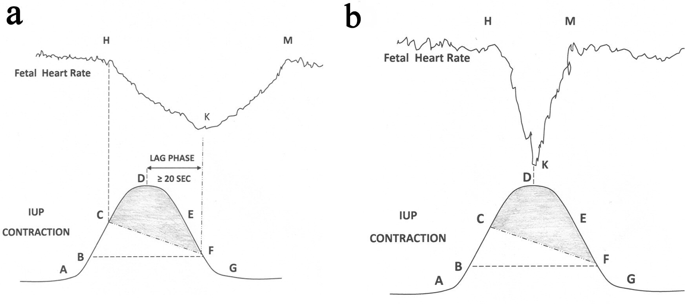
Figure 3. (a) Schematic drawing of FHR deceleration resulting from peripheral chemoreflex due to hypoxemia based on scientific rationale. Hypoxemic trigger is very likely to produce a classical “late deceleration” [9]. Shaded area: level of IUP where fetal PaO2 will continue to drop during deceleration; Point A: contraction commences; B: IUP enough to commence fetal hypoxemia; C: worsening fetal hypoxemia enough to start FHR deceleration; D: peak of contraction where speed of worsening of hypoxemia will slow down but hypoxemia will continue to worsen (PaO2 continues to drop); E: hypoxemia will continue to worsen; F: hypoxia will start recovering because IUP equivalent to point B. Chemoreflex induced FHR deceleration will start recovering at point F and recovery will extend beyond the end of contraction. FHR: fetal heart rate; IUP: intrauterine pressure; PaO2: fetal partial pressure of oxygen. (b): Schematic drawing showing that the common rapid short-lasting FHR decelerations in labor cannot be explained by fetal hypoxemia.


