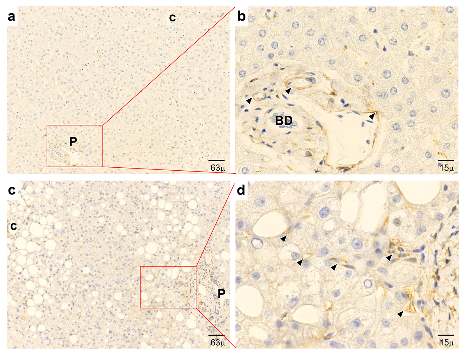
Figure 1. Immunohistochemical expression of APJ. (a, b) In normal control liver, APJ shows positive staining at vascular regions and capillaries in the periportal area. (c, d) In early-stage NASH liver, APJ shows strong positive staining at hepatic sinusoidal lining cells and inflammatory cells in the pericentral area and positive staining at hepatic sinusoidal lining cells in the periportal area. P: portal tract; c: central vein; BD: bile duct. (a, c) Low magnification. (b, d) High magnification.
