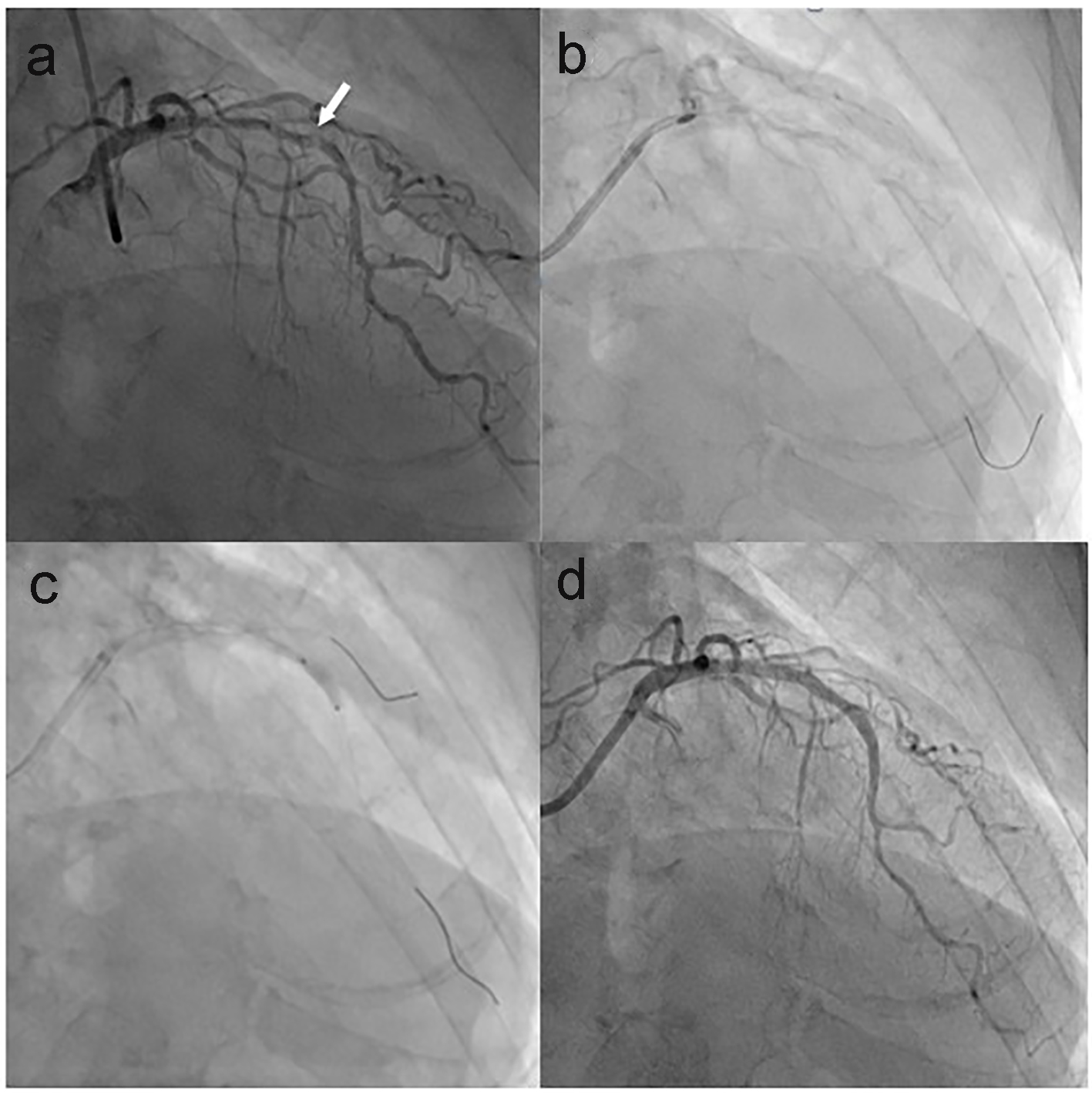
Figure 1. CAG images of the first PCI. (a) Coronary stenosis in the proximal-mid portion of LAD with a severe focal stenosis (arrow). (b) RA with a 1.5-mm burr. (c) Pre-dilation with a 2.5-mm non-compliant balloon. (d) After DES implantation. CAG: coronary angiogram; PCI: percutaneous coronary intervention; LAD: left anterior descending coronary artery; RA: rotational atherectomy; DES: drug-eluting stent.
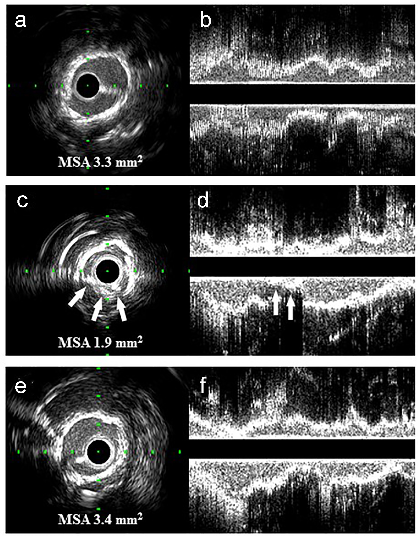
Figure 2. Transversal (a, c and e) and longitudinal (b, d and f) IVUS images of this case: (a, b) immediately after the first PCI; (c, d) the deformed stent struts (arrows) and heavily calcified coronary plaque at the ISR site with an MSA of 1.9 mm2; (e, f) well-expanded ISR site and disappearance of part of the stent struts. IVUS: intravascular ultrasound; PCI: percutaneous coronary intervention; ISR: in-stent restenosis; MSA: minimum stent area.
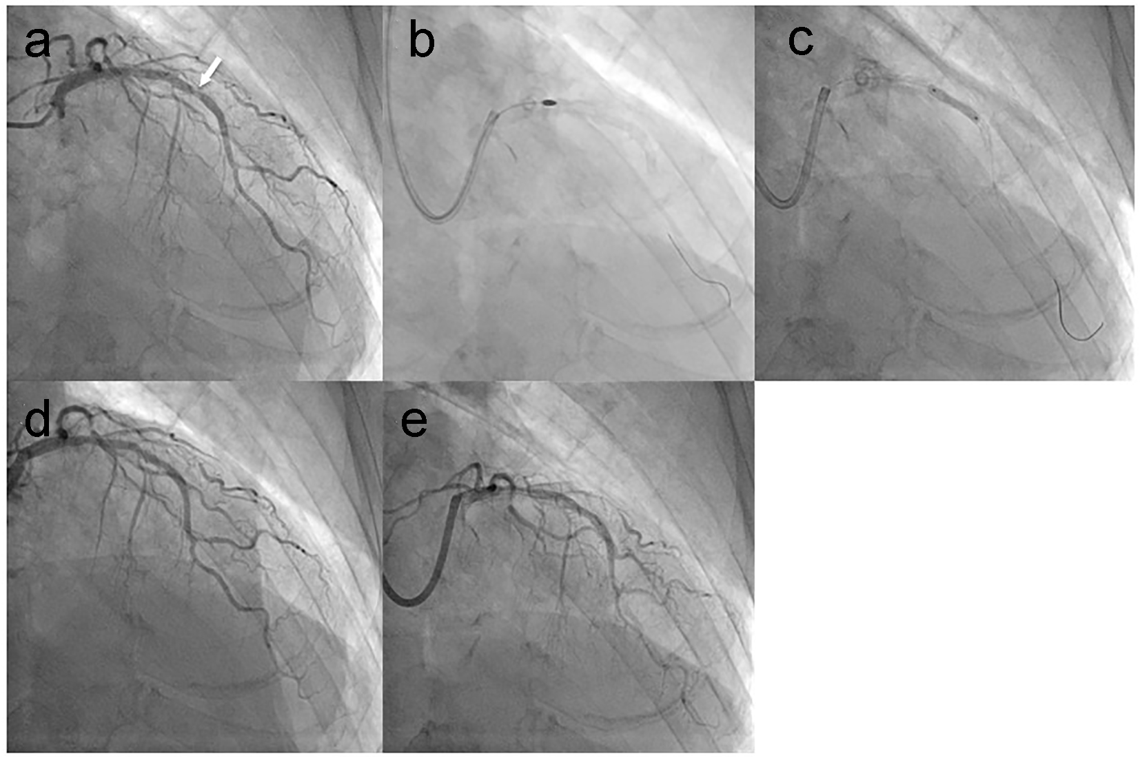
Figure 3. CAG images at 2 months after the first PCI and at follow-up after the final PCI: (a) focal ISR in the mid portion of the LAD (arrow); (b) RA with a 2.0-mm burr; (c) high-pressure dilation with a 2.5-mm non-compliant balloon; (d) immediately after the second PCI; (e) at follow-up after the final PCI. CAG: coronary angiogram; PCI: percutaneous coronary intervention; ISR: in-stent restenosis; LAD: left anterior descending coronary artery; RA: rotational atherectomy.