Journal of Clinical Medicine Research, ISSN 1918-3003 print, 1918-3011 online, Open Access
Article copyright, the authors; Journal compilation copyright, J Clin Med Res and Elmer Press Inc
Journal website http://www.jocmr.org

Case Report

Volume 9, Number 3, March 2017, pages 216-220


Atypical Teratoid/Rhabdoid Tumor of the Sellar Region in an Adult With Long Survival: Case Report and Review of the Literature

Figures

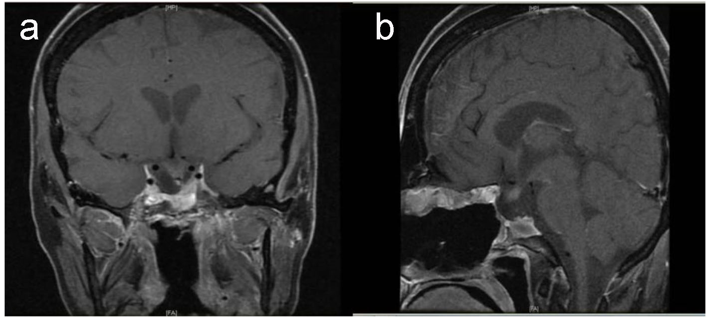
Figure 1.
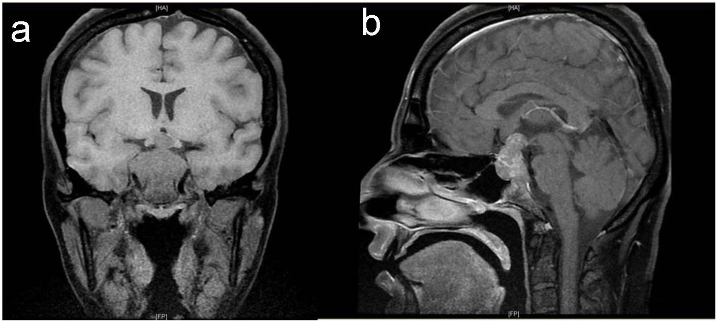
Figure 1. Baseline pituitary MRI. T1-weighted MRI coronal (a) and sagittal (b) images show large intrasellar mass with suprasellar extension compressing and displacing the optic chiasm with evidence of tumor ischemia, bilateral invasion of cavernous sinus and clivus with posterior destruction of clinoid.
Figure 2.
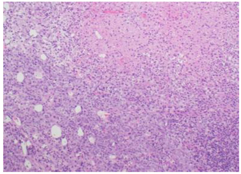
Figure 2. Tumor is composed of sheets of undifferentiated cells with large area of necrosis (H&E stain, ×100 magnifications).
Figure 3.
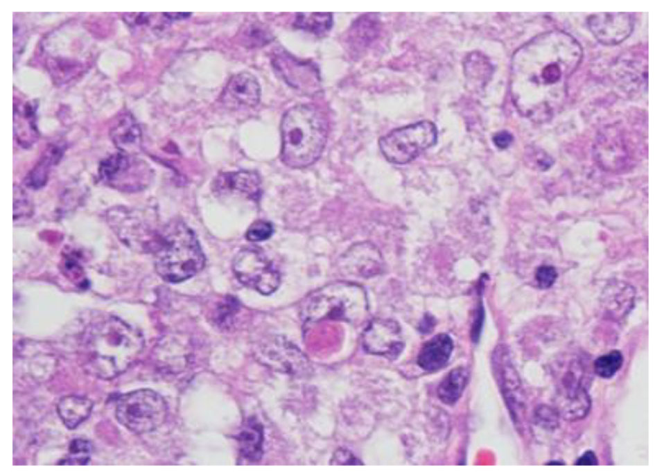
Figure 3. Tumor cells have oval nuclei and prominent nucleoli with focal eosinophilic globular inclusions (H&E stain, × 1,000 magnification with oil).
Figure 4.
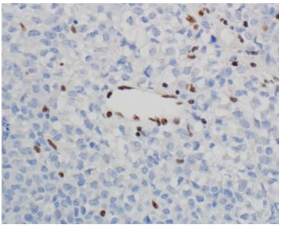
Figure 4. INI-1 (BAF47) immunostain shows loss of nuclear staining in the tumor nuclei and retention of nuclear staining in the lymphocytes and endothelial cells (× 400 magnification).
Figure 5.
Figure 5. Postoperative pituitary. T1-weighted MRI coronal (a) and sagittal (b) images show small residual tumor affecting left side of sella with evidence of empty sella, stable displaced pituitary stalk and down displaced optic chiasm.
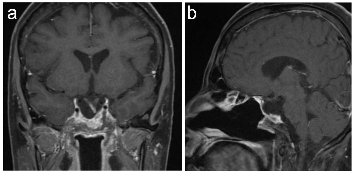
Figure 6.
Figure 6. Latest pituitary MRI. T1-weighted MRI coronal (a) and sagittal (b) images show stable empty sella. Stable deformed left-sided displaced pituitary stalk as well as of down displacement of the optic chiasm and the floor of the third ventricle with focal enhancement of the optic chiasm remains unchanged since the previous study.

Tables

Table 1. Serial Pituitary Hormone Profile
 
DateFSH (IU/L)LH (IU/L)Prolactin (mL/L) (102 - 496)Cortisol (nmol/L)TSH (mIU/L) (0.27 - 4.2)ACTH (pmol/L) (1.6 - 13.9)FT4 (pmol/L) (12.0 - 22.0)
December 2013 (baseline)0.50.235016< 0.0050.18
January 20140.4< 0.1121233< 0.005-15.6
May 20140.5< 0.11337190.024< 0.2211.7
August 20150.2< 0.1263420.25< 0.2210.7
November 20160.2< 0.124060.0090.317.2


Table 2. Summary of Previously Described Cases of Sellar AT/RT and the Present Case
 
AgeSexPresentationTreatmentOutcomeReference
20FVision lossResection, radiation, and chemotherapyAlive at 28 months[11]
31FNot describedResection and radiationDied at 9 months[11]
56FHeadache and diplopiaResection and radiationDied at 23 months[12]
61FSixth cranial nerve palsyResectionDied at 3 months[13]
57FHeadache, diplopia, third CN palsyResection, radiation, and chemotherapyAlive at 6 months[13]
44FVisual disturbanceResection, radiation, and chemotherapyDied at 17 months[14]
60FHeadache and diplopiaResection, radiation, and chemotherapyDied at 30 months[15]
46FHeadacheNot describedNot described[16]
43FHeadache and diplopiaResection and radiationAlive at 2 weeks[17]
36FHeadache and blurred visionResection, radiation, and chemotherapyDied at 29 months[10]
35FHeadache, diplopia, and amenorrheaResection, radiation, and chemotherapyAlive at 37 monthsPresent case