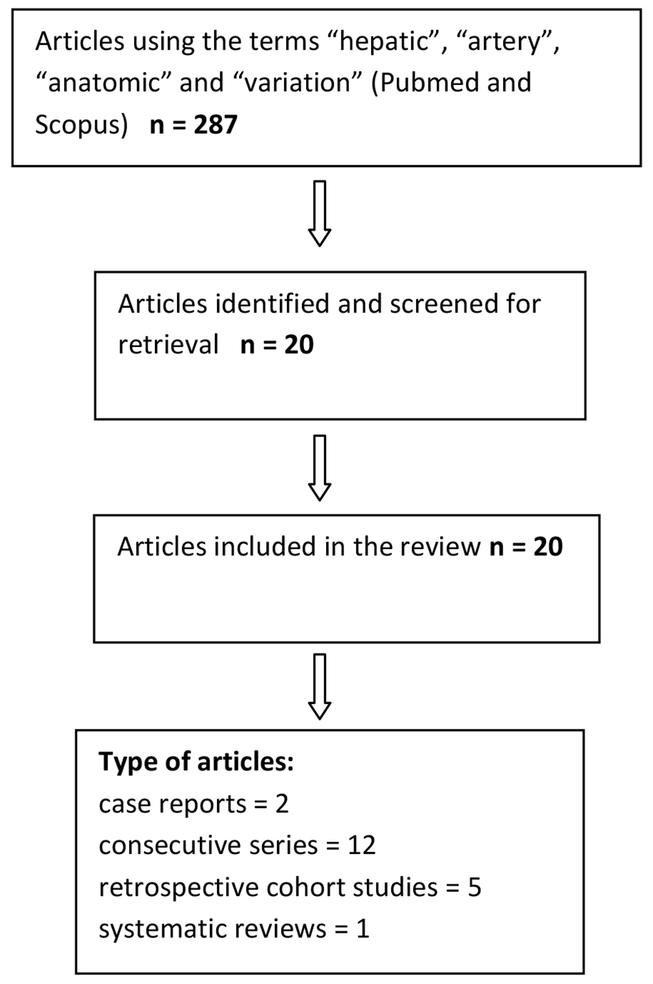
Figure 1. Summary of the literature search.
| Journal of Clinical Medicine Research, ISSN 1918-3003 print, 1918-3011 online, Open Access |
| Article copyright, the authors; Journal compilation copyright, J Clin Med Res and Elmer Press Inc |
| Journal website http://www.jocmr.org |
Review
Volume 9, Number 4, April 2017, pages 248-252
The Main Anatomic Variations of the Hepatic Artery and Their Importance in Surgical Practice: Review of the Literature
Figures
Tables
| Author | No. of patients | Type of patients |
|---|---|---|
| Paraskevas and Raikos [8] | 1 | Cadaver |
| Prakash et al [6] | 50 | Cadaver |
| Chitra [9] | 50 | Cadaver |
| Venieratos et al [7] | 77 | Cadaver |
| Le Bian et al [10] | 2 | Living patient |
| Panagouli et al [11] | 12,196 | Living patient/cadaver |
| Schwartz et al [12] | 1 | Living patient |
| Sureka et al [13] | 600 | Living patient |
| Chaib et al [14] | 60 | Cadaver |
| Ramanadham et al [15] | 2 | Living patient |
| Yi et al [16] | 1 | Living patient |
| Loschner et al [17] | 1,297 | Living patient |
| Lopez-Andujar et al [18] | 1,081 | Cadaver |
| Koops et al [19] | 604 | Living patient |
| De Cecco et al [20] | 250 | Living patient |
| Coskun et al [21] | 48 | Living patient |
| Stemmler et al [22] | 63 | Living patient |
| Winston et al [23] | 371 | Living patient |
| Covey et al [24] | 600 | Living patient |
| Saba and Mallarini [25] | 1,629 | Living patient |
| Sebben et al [26] | 30 | Cadaver |
| Michels type | N = 19,013 | 100% |
|---|---|---|
| I | 15,342 | 81 |
| II | 556 | 3 |
| III | 710 | 3.7 |
| IV | 163 | 0.8 |
| V | 592 | 3.2 |
| VI | 309 | 1.6 |
| VII | 38 | 0.2 |
| VIII | 66 | 0.35 |
| IX | 245 | 1.2 |
| X | 5 | 0.04 |
| Not yet classified types | 784 | 4.1 |