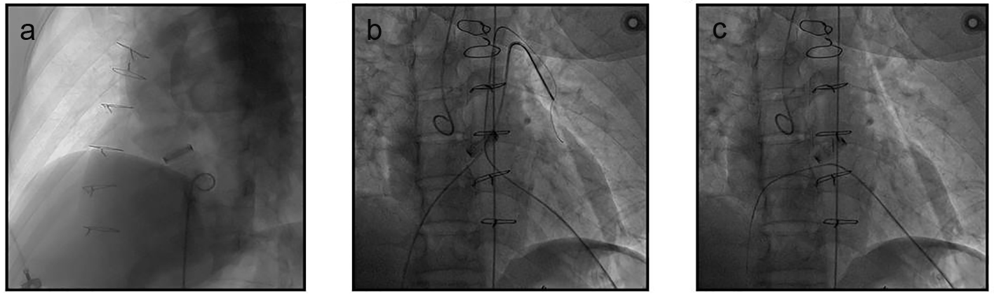
Figure 1. Angiography assessment and the establishment of the apex-venous loop. (a) LV angiography confirmed the membranous VSD. (b, c) Through the apex, a guide wire was advanced across the VSD to the right ventricle and then to the left pulmonary artery where it was snared (b) and exteriorized from the RFV (c), thus the apex-venous loop was established.
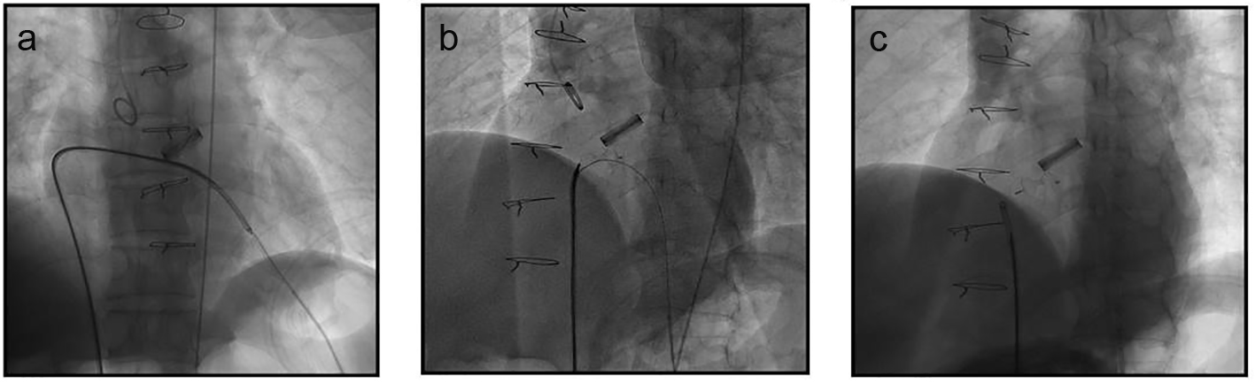
Figure 2. Occluder deployment through the apex-venous loop. (a) Occluder deployment was performed with the distal tip of the delivery sheath pointing to the apex. (b) The left disk was carefully opened and positioned beneath the edge of mechanical valve followed by the opening of the waist and the right side of the occluder. (c) The occluder was released and the loop wire was withdrawn after final LV angiography confirmation.