Journal of Clinical Medicine Research, ISSN 1918-3003 print, 1918-3011 online, Open Access
Article copyright, the authors; Journal compilation copyright, J Clin Med Res and Elmer Press Inc
Journal website http://www.jocmr.org

Case Report

Volume 8, Number 10, October 2016, pages 760-763


Unusual Survival of Anomalous Left Coronary Artery From the Pulmonary Artery With Severe Rheumatic Mitral Stenosis in Septuagenarian Women: Foes Becoming Friends?

Figures

Figure 1.
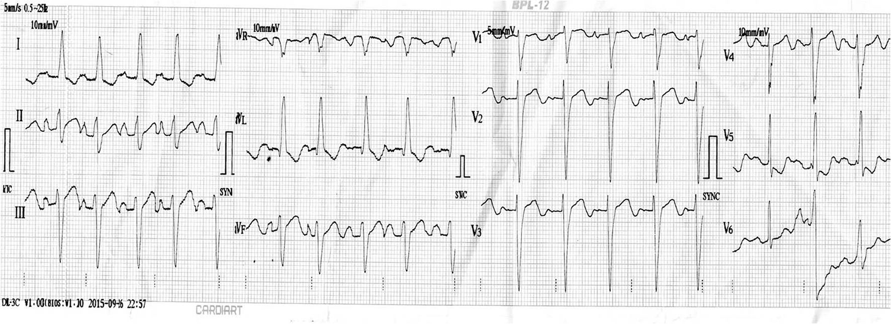
Figure 1. ECG displaying normal sinus rhythm, biatrial enlargement, left axis deviation, poor R progression and ST-T changes in leads V5-6.
Figure 2.
Figure 2. Chest skiagram PA view displaying full pulmonary bay and dilated right pulmonary artery along with features of pulmonary congestion.
Figure 3.
Figure 3. Trans-thoracic echocardiogram and color Doppler showing severe mitral stenosis (A, B), mild mitral regurgitation and interventricular septal fluttering (white arrow, C), and dilated right coronary artery and left coronary artery originating from main pulmonary artery (D).
Figure 4.
Figure 4. Continuous flow entering the pulmonary trunk.
Figure 5.
Figure 5. Coronary angiogram showing absence of a left coronary ostium in aortic sinus (white arrow head; A), dilated and tortuous right coronary artery (red arrow; B), intercoronary collaterals communicating with left coronary artery (white arrows; C), and origin of left anterior descending and left circumflex coronary arteries from the main pulmonary artery on reconstructed coronary CT angiogram (D).