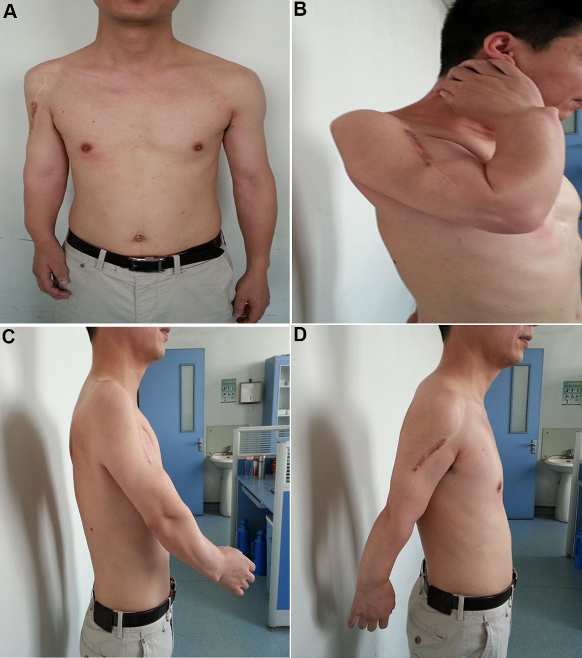
Figure 1. Radiographic evaluation of the surgical treatment at pre- and postoperative stages. (A and B) Radiograph showing cartilaginous lesions or enchondromas predominantly localized in the upper part of the humerus (A) and the lower part of the radius (B) of the 16 years old boy affected with Ollier disease. (C) A huge tumor of the proximal humerus with massive cortical destruction and soft tissue extension was noticed at age 25 of the patient. (D) The radiograph on the postoperative day 2 (anteroposterior view). The prosthesis was well in place after the surgery. (E) The radiograph on 1 year after the surgery (anteroposterior view). The prosthesis was still well in place. (F) The radiograph on 8 years after the surgery (anteroposterior view). The prosthesis remained well in place. (G and H) The radiographs of the ulna and radius of the limb were also taken at age 33 of the patient (8 years after the surgery). There were no signs for malignant transformation in those lesions.