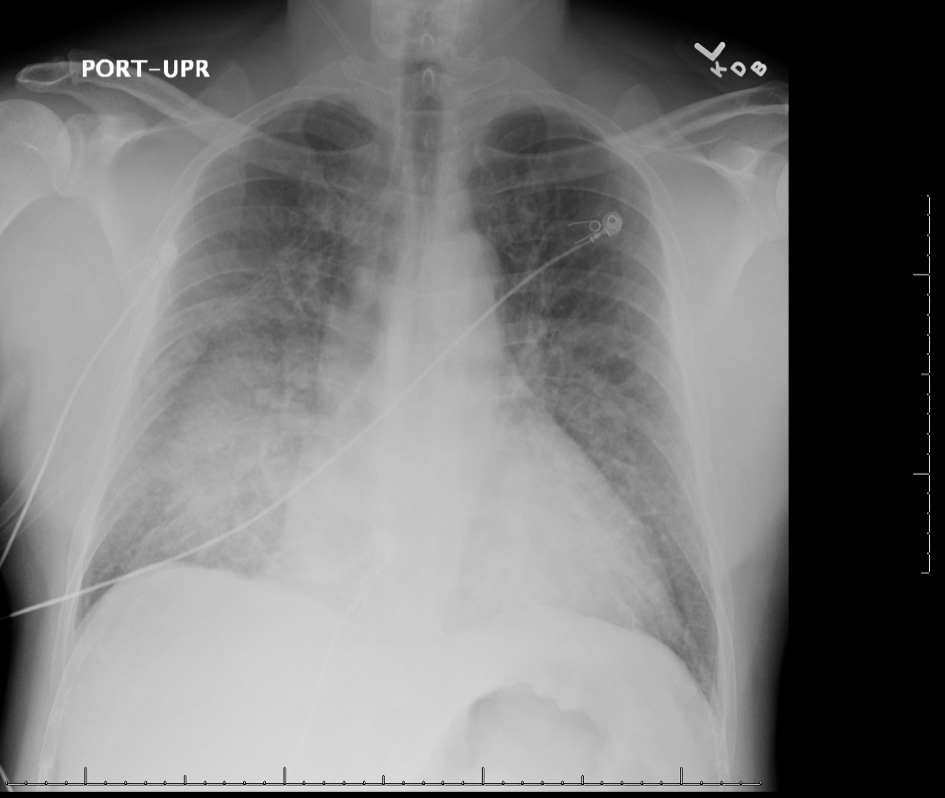
Figure 1. Chest X-ray (anterior-posterior view) showing cardiomegaly, perihilar consolidations, and suspected pulmonary edema with trace effusions (scale on screen 1 cm increments).
| Journal of Clinical Medicine Research, ISSN 1918-3003 print, 1918-3011 online, Open Access |
| Article copyright, the authors; Journal compilation copyright, J Clin Med Res and Elmer Press Inc |
| Journal website https://www.jocmr.org |
Case Report
Volume 15, Number 6, June 2023, pages 332-335
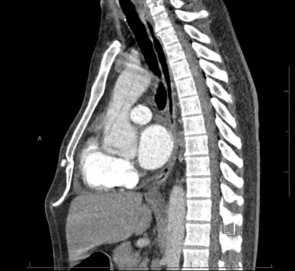
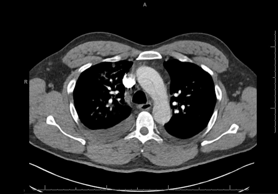
An Uncommon Presentation of Acute Thoracic Aortic Dissection
Figures