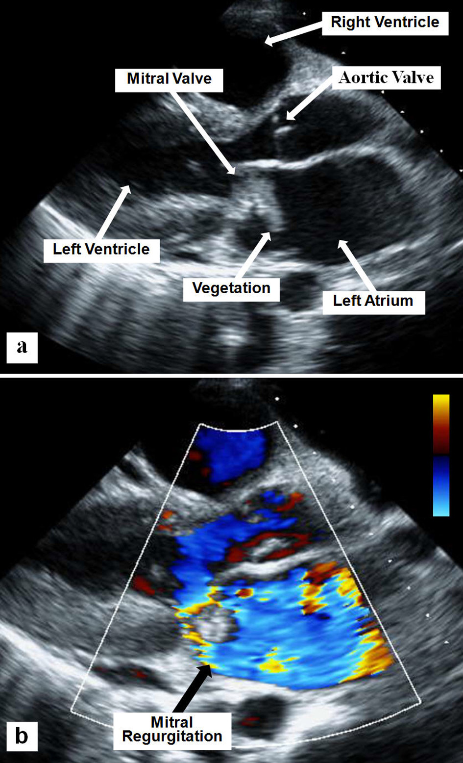
Figure 1. Two-D parasternal long axis echocardiographic view showing the mitral valve vegetation (a) and the severe mitral regurgitation using superimposed color flow Doppler (b).
| Journal of Clinical Medicine Research, ISSN 1918-3003 print, 1918-3011 online, Open Access |
| Article copyright, the authors; Journal compilation copyright, J Clin Med Res and Elmer Press Inc |
| Journal website http://www.jocmr.org |
Case Report
Volume 7, Number 6, June 2015, pages 485-489
Iatrogenic Aortic Insufficiency Following Mitral Valve Replacement: Case Report and Review of the Literature
Figures
Table
| Non-coronary cusp | Right coronary cusp | Left coronary cusp | |
|---|---|---|---|
| MVA: mitral valve annuloplasty ring; MVR: mitral valve replacement. | |||
| Hill et al [1] | MVR (two cases) | VSD repair | MVR |
| ASD repair | |||
| VSD repair | |||
| Ducharme et al [2] | MVA | ||
| Rother et al [3] | MVA | ||
| Aboelnasr and Rohn [4] | MVA | ||
| Mehta and Hunsaker [5] | MVA | ||
| Santiago et al [6] | MVR | MVA | |
| Dogan et al [7] | MVR | ||
| Al Yamani et al [8] | MVR | ||
| Oakley et al [9] | MVR | ||
| Dreyfus et al [10] | MVA | ||
| Pagel et al [11] | MVA | ||
| Rey et al [12] | ASD repair (nine cases) | ||
| Woo et al [13] | VSD repair | ||